
When “Restless Legs” Was Really a Fascial Adhesion
A calf-pain case report: four years, one back surgery, and a return to 25 miles a week.
A 48-year-old recreational distance runner arrived at our Los Angeles clinic after four years of deep right-calf pain that was worst at night, in bed, the moment she lay still. Along the way she had been told it might be a pinched nerve in her back — and even had spine surgery for it — while others wondered about restless legs. Her vascular studies were normal. Her nerve conduction study was normal. What finally explained it was a small fascial adhesion we could see on ultrasound — and release.
Editor’s note: shared for education. Identifying details — name, exact age, occupation, and the specifics of prior care — have been changed or generalized to protect privacy. “Dana” is a pseudonym. This describes one patient’s course; it is not individual medical advice, and no specific outcome or cure is implied.
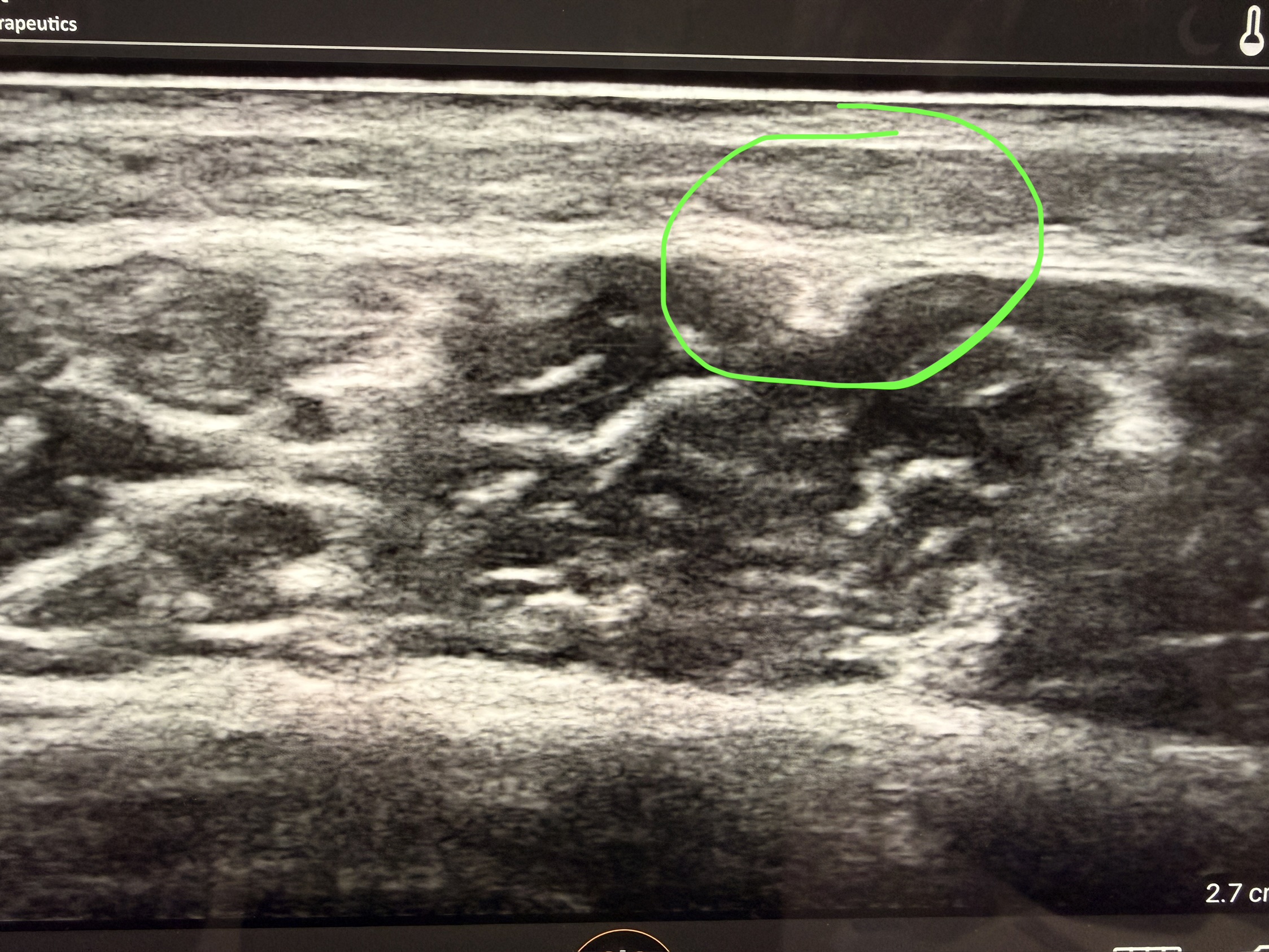
Calf fascial adhesion
A four-year problem with a five-minute imaging answer
Dana, a 48-year-old woman who had run for most of her adult life, developed a deep ache and tightness in her right calf. It was quiet during the day and loud at night: as soon as she lay down to sleep, the leg would start to gnaw and she felt a restless urge to move it — symptoms that looked, on the surface, like restless legs syndrome. Over four years she saw several clinicians. A workup pointed toward lumbar nerve involvement, and she ultimately underwent lumbar spine surgery for a presumed radiculopathy. Her calf pain did not change. A vascular workup — arterial and venous studies — was normal. A nerve conduction study (EMG/NCS) was normal. When she came to us, we did not order another scan first; we put an ultrasound probe on the calf that hurt — over the exact spot that was tender when we pressed on it. There, in the superficial fascial layer of the gastrocnemius, was a thickened, tethered adhesion. We released it with Hydrofascia Release. Her pain resolved completely and stayed gone, and she returned to running about 25 miles a week.
The four-year detour
Chronic calf pain is deceptively hard to sort out, because so many different problems can produce it and the obvious ones get ruled in first. Dana’s story followed the usual map:
Every one of those tests did its job. The gap was not a missed reading — it was that none of them image the fascia, and fascia was where the problem lived. This is a recurring theme in our clinic and the reason we so often start with a dynamic ultrasound of the tissue that hurts.
Why a fascial adhesion feels worse when you lie down
The most confusing part of Dana’s story was the timing. Most musculoskeletal pain is worse with activity and better with rest. Hers was the opposite — comfortable on the run, miserable in bed. That reversal is exactly why she kept being pointed toward restless legs. Here is the working explanation we gave her, offered as clinical reasoning rather than proven fact:
When you stand and walk, your calf muscles act as a pump. Every step squeezes the deep veins and moves blood and fluid up and out of the leg, and the muscle layers glide over one another to do it. When you lie down at night, that pump switches off. Blood and fluid redistribute, pressures in the leg shift, and the tissues settle. In a normal calf, none of this is noticeable. But where a fascial layer is stuck to the one beneath it, that adhesion tethers small nerves and vessels and resists the change. As flow shifts away from and around the restricted area at rest, the tethered tissue becomes irritable — producing a deep, crawling ache and an urge to move. Moving, of course, turns the pump back on and briefly relieves it. The result is a peripheral, one-sided problem that impersonates a central one.
The clinical lesson is not that restless legs syndrome isn’t real — it very much is, and it deserves proper neurological workup.4 The lesson is that “worse at rest, better with movement, relieved when I get up” can also be the signature of a mechanical fascial problem, especially when it is one-sided, tied to a specific spot, and unresponsive to the treatments RLS usually responds to. In a runner, chronic calf pain also overlaps with chronic exertional compartment syndrome, another condition where the fascia — not the muscle, nerve, or artery — is the culprit.5
What the ultrasound found that the other tests couldn’t
We examined the symptomatic calf with high-resolution, dynamic ultrasound — imaging the tissue while it moves, not just at rest — and focused on the point that was tender to palpation. In the superficial fascial layer of the gastrocnemius, where the tissue should glide smoothly, was a thickened, tethered plane that did not slide when the ankle was moved. That is a fascial adhesion (Figure 1), and it sat directly beneath the spot that reproduced her pain on exam.
This is precisely the kind of change static imaging tends to miss. MRI is superb for tears, tumors, and bone, but a thin, non-gliding fascial layer can look unremarkable on a still image. Ultrasound, by contrast, can show altered connective-tissue structure and, uniquely, can watch tissue move in real time — which is why researchers have used it to document abnormal fascial thickness and shear in people with chronic pain.6 A detailed, head-to-toe version of this same approach is what we call Fascial Mapping, and the “normal scans but real pain” story it so often uncovers is one we’ve written up before.
Hydrofascia Release: separating what shouldn’t be stuck
Hydrofascia Release (ultrasound-guided hydrodissection) treats an adhesion directly. Under live ultrasound, a small volume of fluid is placed precisely into the stuck fascial plane. The fluid does mechanically what the tissue can no longer do on its own: it separates the layers, frees the tethered nerves and vessels, and restores glide. It is not a cortisone shot and it is not surgery — it is a targeted, image-guided release of the exact interface that isn’t moving.
This technique is well described in the sports-medicine literature and is part of our founder’s own published work. Dr. Jacques Courseault first described ultrasound-guided fascial hydrodissection for chronic hamstring injury,2 and a separate published case documented fascial hydrodissection resolving chronic calf pain in a former marathon runner whose imaging, like Dana’s, was normal — and who returned to running.3 For runners whose calf problem is a chronic exertional compartment issue, our team has also reported resolving symptoms with ultrasound-guided hydrodissection combined with dextrose prolotherapy — a non-surgical alternative to fasciotomy.1 Where an adhesion sits alongside instability or a stubborn tendon, we may add prolotherapy or ProloBoost PRP; where the goal is durable return to sport, we bridge into the Fascia Performance Program.
Complete relief — and it held
Dana reported complete resolution of her calf pain — a full 100% improvement by her own account — including the nightly restlessness that had disrupted her sleep for four years. At follow-up the improvement was maintained. She has returned to running roughly 25 miles per week without pain, and without the nightly urge to move her leg. After four years, three normal tests, and a spine surgery that never addressed the real problem, the fix was releasing a single fascial adhesion the other studies could not see.
Individual results vary. This is a single de-identified case and is not a promise of a particular outcome. A careful evaluation is the only way to know whether a fascial cause is contributing to a given person’s pain.
Two patterns worth a fascial evaluation
Dana’s case sits at the crossing point of two situations we see constantly. If either sounds familiar, an ultrasound of the tissue that hurts is a reasonable next step.
Leg pain or an urge to move that is worst lying down or at night, eases when you get up, and hasn’t responded to the usual restless-legs measures — especially if it is one-sided or tied to a specific spot.
Chronic calf, leg, or muscle pain — often in a runner or athlete — with normal MRI, EMG, and vascular studies, and a surgery or injection that didn’t fix it. Fascia is the one tissue those tests don’t really evaluate.
Takeaways for clinicians and patients
Written for The Fascia Institute and Treatment Center of LA. Clinical approach directed by Dr. Jacques Courseault, MD, founder of the Fascia Institute and a physician who has published on ultrasound-guided fascial hydrodissection.
Calf pain, restless legs & fascia
How do I know if my leg pain is restless legs syndrome or something else?
True restless legs syndrome is usually a symmetrical, whole-limb urge to move with well-described neurological features, and it responds to specific treatments. Pain that is one-sided, focused on a particular spot, tied to an old injury or heavy training, and unresponsive to restless-legs measures is worth evaluating for a mechanical or fascial cause. Restless legs has a long differential and is commonly over-diagnosed, so a careful workup matters.4
My MRI, EMG, and vascular tests were all normal. Why do I still hurt?
Those tests are excellent at what they check — discs and nerve roots, nerve conduction, and blood flow — but none of them really evaluate fascia, the connective-tissue layers that let muscles glide. An adhesion between layers can cause real, persistent pain while every standard test reads normal. A dynamic ultrasound of the tissue that hurts is what closes that gap.
What is Hydrofascia Release and is it surgery?
It is a non-surgical, in-office procedure. Under live ultrasound, a small amount of fluid is placed into a stuck fascial plane to separate the layers and free tethered nerves and vessels — restoring glide. It is not a steroid injection and it does not require an incision. Learn more on our Hydrofascia Release page.
I’m a runner with chronic calf pain that won’t quit. Could this help me?
Possibly. Chronic calf pain in runners has several causes, including fascial adhesions and chronic exertional compartment syndrome — both fascia-driven and both often missed by static imaging.5 Published cases describe fascial hydrodissection helping runners return to sport after years of failed conservative care.3 An evaluation is the way to find out whether it applies to you.
Do you see patients from outside Los Angeles?
Yes. Our clinic is in Beverly Hills and we see local and traveling patients. For those in California, some visits can be handled by telehealth, though hands-on ultrasound evaluation and procedures are done in-clinic. You can request an appointment or see pricing.
Still chasing a pain no scan can explain?
If your leg pain is worse at night, your tests are normal, or a treatment didn’t hold, we can put an ultrasound on the tissue that hurts and look for a fascial cause. Beverly Hills · serving greater Los Angeles.
415 North Crescent Drive, Suite 130, Beverly Hills, CA 90210 · fit@fasciala.com
Selected literature
- Cristian AL, Courseault J. Successful Treatment of Chronic Exertional Compartment Syndrome with Hydrodissection & Prolotherapy. Association of Academic Physiatrists, Physiatry ’24 Annual Meeting; Poster 1028; Feb 23, 2024. Abstract.
- Courseault J, Kessler E, Moran A, Labbe A. Fascial Hydrodissection for Chronic Hamstring Injury. Curr Sports Med Rep. 2019;18(11):416–420. PMID: 31702723. doi:10.1249/JSR.0000000000000650.
- Bailowitz Z, Chang CJ. Ultrasound-guided Fascial Plane Hydrodissection for Chronic Calf Pain. PM R. 2021;13(S1). Abstract.
- Kwatra V, Khan MA, Quadri SA, Cook TS. Differential Diagnosis and Treatment of Restless Legs Syndrome: A Literature Review. Cureus. 2018;10(9):e3297. PMID: 30443467. doi:10.7759/cureus.3297.
- Tarabishi MM, Almigdad A, Almonaie S, Farr S, Mansfield C. Chronic Exertional Compartment Syndrome in Athletes: An Overview of the Current Literature. Cureus. 2023;15(10):e47797. PMID: 38022185. doi:10.7759/cureus.47797.
- Langevin HM, Stevens-Tuttle D, Fox JR, et al. Ultrasound evidence of altered lumbar connective tissue structure in human subjects with chronic low back pain. BMC Musculoskelet Disord. 2009;10:151. PMID: 19958536. doi:10.1186/1471-2474-10-151.
The Fascia Institute and Treatment Center operates a connected network. On the West Coast, this Beverly Hills clinic serves greater Los Angeles. Our New Orleans flagship, fasciainstitute.org, is where founder Dr. Jacques Courseault develops and publishes this work — including a review of dextrose prolotherapy for lower-extremity injuries.
Because many hard-to-explain, body-wide pain patterns trace back to connective-tissue laxity, our dedicated Hypermobility & Ehlers-Danlos Clinic and its EDS blog go deeper on that side of the picture.
This case report is provided for general educational purposes and is de-identified. It describes the course of a single patient; individual results vary and no specific outcome or cure is implied. Nothing here is a substitute for evaluation and advice from your own qualified healthcare provider. If you have sudden, severe, or rapidly worsening leg pain, swelling, numbness, or discoloration, seek urgent medical care to rule out a clot or acute vascular problem.


